SB Cells®
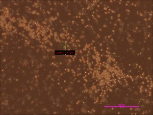
Our stem cells are mainly from human adult blood and bone marrow. They do exist in other tissues and organs; however, blood and bone marrow are relatively easy to obtain compared to other samples. After receiving blood and bone marrow, we separate the cells into two distinct groups. The first group contains larger cells such as white cells and red blood cells while the other group contains our stem cells. We have dubbed these cells SB Cells® (StemBios cells). There are two groups of SB Cells® : SB-1 and SB-2 (Figure 1). We discovered that SB Cells® in general also contain positive markers for Nanog and Oct 4. Since Nanog and Oct 4 are markers for Embryonic Stem Cells, the expression of Nanog and Oct 4 on SB Cells® leads us to believe that SB Cells® have the characteristics of Embryonic Stem Cells and are relatively small in size.
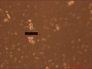
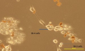
After one to two weeks of media incubation in vitro, the size of the SB Cells® increases significantly. We call these larger cells SB-3 cells (Figure 2). More importantly, SB-3 cells can be expanded into a larger population. Once the SB-3 cells attach to the dish, we call them SB-4 cells (Figure 3), having grown even larger in size. SB-4 cells can undergo at least 40 passages with normal karyotype and normal telomerase activity. Its doubling time is approximately 24 hours. When growth factor for ectoderm is added, SB-4 cells differentiate into dopaminergic neuron, which is associated with Parkinson’s disease.
When growth factor for endoderm is added, SB-4 cells differentiate into liver cells and will express albumin, which can be used as a carrier for small pills/drugs. SB-4 cells can also differentiate into islet cells. These cells are believed to be an important element for curing diabetes.
SB Cells® can differentiate into different types of mesoderm cells including, but not limited to, osteocyte, chondrocyte, adipocyte, and muscle cells (including cardiomyocyte). These have the potential to cure cardiovascular-related diseases, the number one cause of death in America. Recently, StemBios has undergone in vivo cell tracking in mice in addition to our in vitro studies yielding results that highly suggest VSEL (very small embryonic-like stem cell; CD133), MSC (Mesenchymal stem cell; Stro 1) and BLSC (Blastomere-like stem cell; CEA) are downstream from SB Cells® .

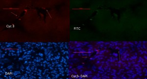
Figure 4. In vivo tracking of Human male SB Cells® in the female SCID mouse liver: Injection of SB Cells® staining with fish y-Cyc3 chromosome
In other words, our SB Cells® are much closer to Embryonic Stem Cells than VSEL, MSC and BLSC. We, therefore, believe SB Cells® have the potential to differentiate into other body cells similar to Embryonic Stem Cells. This hypothesis has also been demonstrated by our in vivo cell tracking assay where our human SB Cells® were injected into mice to observe differentiation in these mice into neurons (ectoderm), liver cells (endoderm) (Figure 4) and muscle cells (mesoderm). More importantly, unlike Embryonic Stem Cells, our SB Cells® will not form Teratoma and will not cause immune-mediated rejection as long as it is autologous.


